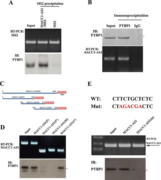
Fig. 5. Identification of PTBP1 as a MACC1-AS1-binding partner.
a PTBP1 co-precipitated with MACC1-AS1. Upper: MACC1-AS1-MS2-pulldown assays, MS2 was used as a control. Lower: western blot analysis of the presence of PTBP1 in the MACC1-AS1-MS2 precipitates. b MACC1-AS1 was detected in the PTBP1 IP. Upper: western blots of PTBP1 in the IP, normal IgG was used as a control. Lower: RT-PCR and agarose gel electrophoresis indicated the association of MACC1-AS1 with PTBP1. c Schematic representation of truncated MACC1-AS1-MS2 RNAs, which can be bound to amylose resin through MBP-MCP. d Upper: vectors expressing MS2-fused full-length MACC1-AS1 or truncated MACC1-AS1 (5′, middle (m) and 3′) were transiently transfected into HEK-293T cells. RT-PCR showing that individual MS2-fused RNAs were pulled down by MBP-MCP attached amylose resin. Lower: western blots indicating that PTBP1 co-precipitated with the full-length and the 3′ part of MACC1-AS1. e Upper: a putative motif for the PTBP1 binding site within MACC1-AS1 is shown. Red nucleotides indicates the mutated sequences. Lower; wild-type and mutant MACC1-AS1-MS2 RNAs were transfected into HEK-293T cells. MS2-pulldown assays and immunoblots showed that PTBP1 was barely detected in the PTBP1 precipitates when the PTBP1-binding motif within MACC1-AS1 was mutated.

