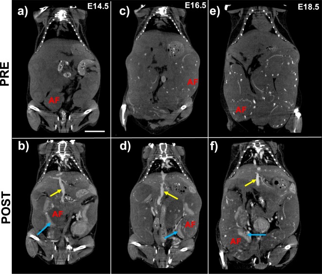
Figure 4.
Nanoparticle contrast-enhanced computed tomography (CE-CT) demonstrates uniform vascular enhancement and enables validation of MRI-derived placental fractional blood volume (FBV). Comparison of pre-contrast and post-contrast coronal images for (a,b) E14.5, (c,d) E16.5, and (e,f) E18.5 show signal enhancement in the IVC (yellow arrow) and placenta (blue arrow). Fetal skeletal features within the amniotic fluid (AF) compartment are visible in both pre-contrast and post-contrast images and become more pronounced at later stages of gestation. Scale bar represents 1 cm.

