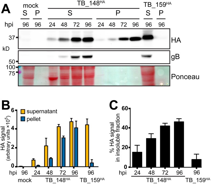
FIG 8.
Solubility analysis of UL148 and Rh159. (A) Human fibroblasts were infected at an MOI of 1 TCID50/cell with TB_148HA, HCMV strain TB40/E-derived virus that expresses UL148 fused to a C-terminal HA tag, or TB_159HA, which lacks UL148 and instead expresses rhesus CMV Rh159 carrying a C-terminal HA tag. At the indicated times postinfection (hpi), infected cells were collected in radioimmunoprecipitation (RIPA) lysis buffer and centrifuged at 21,000 × g for 30 min, after which supernatant (S) and pellet (P) fractions were boiled in gel loading buffer containing 2% sodium dodecyl sulfate (SDS). Equivalent portions of supernatant and pellet were resolved by SDS-PAGE and transferred to a nitrocellulose membrane for detection of protein species immunoreactive to antibodies against HA and gB and for total protein signal using Ponceau S reagent (Ponceau). (B and C) Signal intensity of fluorophore-conjugated secondary antibodies in anti-HA Western blots were measured from three independent biological replicates of the experiment shown in panel A; error bars indicate standard deviations. (B) The fluorescent signal for each infection time point condition (y axis is in arbitrary units, in hundred thousands). (C) The amount of signal found in the insoluble (pellet) fraction relative to the total signal (pellet plus supernatant) for each infection time point is plotted as a percentage value.

