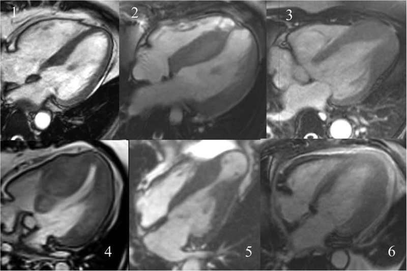
Figure 1. Steady state free precession 4-chamber long axis cine images from individual patients with the 6 different morphologic subtypes of HCM.
The six subtypes shown are: 1) localized basal septal hypertrophy 2) reverse curvature septal hypertrophy 3) apical HCM 4) concentric HCM 5) mid-cavity obstruction with apical aneurysm or 6) other

