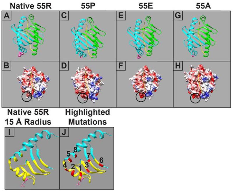
Figure 1:

Molecular modeling of the SLA-DQα*0101 and SLA-DQβ1*0601 heterodimer. Four variants are shown, each having a unique amino acid at position 55 in the beta chain. Upper panels show ribbon models of SLA-DQ with the α chain in green, the DQ β chain in blue, and the peptide in yellow. The side chain of the amino acid 55 is shown in a space-filling representation and colored pink. The lower panels show models of surface electrostatic potential of the various SLA-DQ proteins ranging from −10 kcal/e.u (red) to 0 kcal/e.u (white) and +10 kcal/e.u (blue). The circle highlights the location of amino acid 55. Panels A,B: naturally occurring SLA-DQ molecule with arginine at position 55 in the SLA-DQβ1*0601 chain (55R). Panels C,D: SLA-DQβ1*0601 with position 55 mutated to proline (55P). Panels E,F: SLA-DQβ1*0601 with position 55 mutated to glutamate (55E). Panels G,H: SLA-DQβ1*0601 with position 55 mutated to alanine (55A). Panel I: A ribbon model showing the predicted structure of the natural variant of SLA-DQβ1*0601 chain having arginine at position 55 (pink amino acid with side chain). The yellow portions of the ribbon highlight a region of the protein within 15 Å of amino acid 55. Panel J: Eight amino acid positions, highlighted in red, lie within the 15 Å radius of amino acid 55 and were chosen for mutagenesis studies. The specific amino acid changes at each position are shown in table 2.
