
Figure 1.
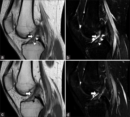
(a) T2 sagittal view: Anterior cruciate ligament showed poor collagen alignment, but still consistent between femur and tibia insertion. Several small enhancements and irregular architecture over anterior cruciate ligament tibial plateau insertion and its main trunk were observed. (b) T2 sagittal view with fat suppression view: Several small enhancements over anterior cruciate ligament tibial plateau insertion. The lesions were diagnosed as the anterior cruciate ligament tear were observed. (c) T2 sagittal view: Decreased enhancements of anterior cruciate ligament tear in tibial plateau area and its main trunk were observed. (d) T2 sagittal view with fat suppression view: Decreased enhancements of anterior cruciate ligament tear in tibial plateau area and its main trunk were observed. Less bony edema was noted. White arrow: Anterior cruciate ligament. White arrowhead: Anterior cruciate ligament tear. Black arrowhead: Healing site of anterior cruciate ligament tear. White small plots: Anterior cruciate ligament border