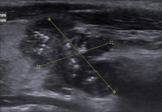
Figure 2.

A 14-year-old boy with American College of Radiology of Radiology Thyroid Imaging Reporting and Data System category 5 nodule. Sagittal gray-scale ultrasonographic image of the left lobe of the thyroid shows a solid markedly hypoechoic mass with lobulated margins and punctate echogenic foci in the lower pole. Histopathological analysis revealed papillary thyroid cancer
