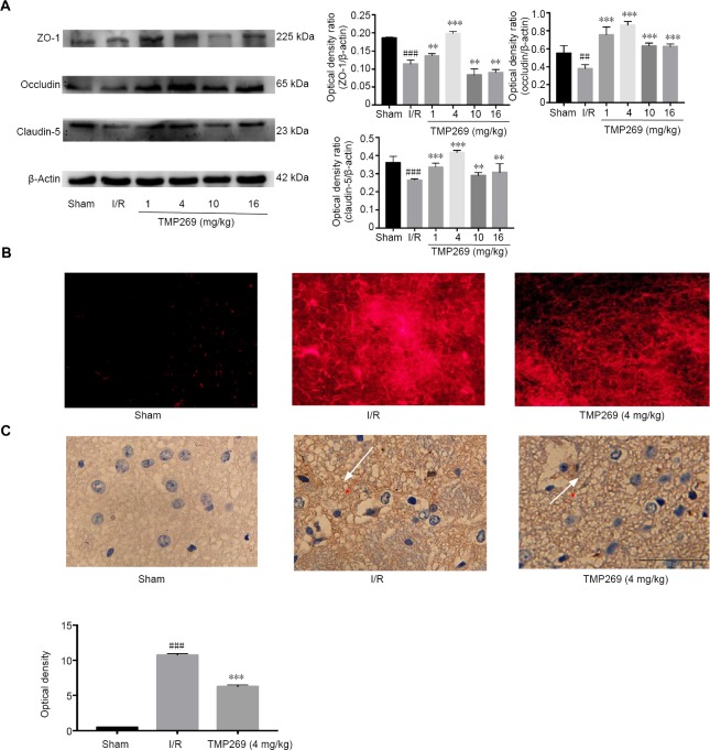
Figure 3.
TMP269 improves I/R-induced abnormal endothelial cell permeability.
(A) Representative western blots and quantified data showing tight-junction protein levels in ischemic rats after 1.5 hours of middle cerebral artery occlusion and 24 hours of reperfusion (I/R). (B) Immunofluorescence images of brain sections after injection of Evans blue compared with the sham group. (C) Immunohistochemistry images and quantitative analysis of mean optical density showing neurofilament staining in different groups. The white arrows indicate the nerve fiber network; IgG leaked into the nerve fiber network of the extravascular space after brain injury. The darker the coloration, the more severe the damage. Scale bar: 100 μm. Data are expressed as the mean ± SD (n = 6). **P < 0.01, ***P < 0.001, vs. I/R group; ##P < 0.01, ###P < 0.001, vs. sham group. I/R: Ischemia/reperfusion.

