. 2019 Nov 14;42(3):560–570. doi: 10.1590/1678-4685-GMB-2018-0148

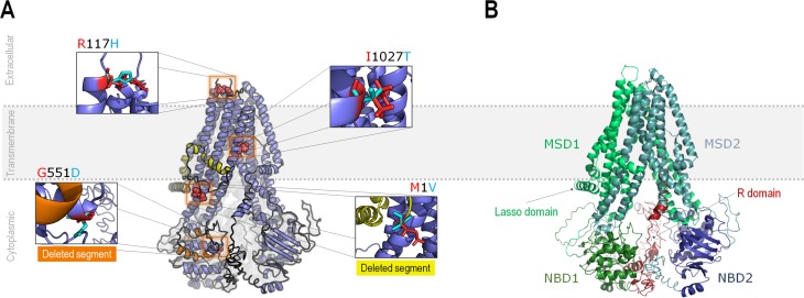
Figure 3. Modeled structure of the CFTR protein. A) Modeled structure for CFTR in its proposed location when inserted in biomembranes (dotted lines), evidencing the absence of extracellular domains. Proposed variants are highlighted, while two segments that may be lost (at least partially) as a function of these variants are highlighted in orange (associated with p.Met1Val) and in yellow (associated with p.Arg117His). Details regarding these deletions are given in the results session. B) Main structural features of human CFTR. The semi-symmetric structure is composed of two equivalent halves, comprising two domains (MSD and NBD) each. The N-terminus half of the protein has exclusive topological regions, namely the Lasso domain and the R domain (or R insertion). MSD: Membrane-spanning domain; NBD: Nucleotide-binding domain; R domain: Regulatory domain.

Figure 3