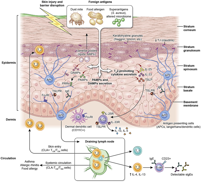
FIG 5.
Skin structure and abnormalities associated with AD. Impaired skin barrier promotes foreign antigen (eg, dust mites and food allergens) penetration and activation of innate immune and pattern recognition receptors. Pathogen-associated molecular patterns and damage-associated molecular patterns are secreted secondary to tissue damage and/or an altered microbial profile to initiate and perpetuate tissue inflammation. Concurrently, antigen stimulation leads to TH2-promoting cytokine secretion (IL-25, IL-33, and TSLP), consequent IgE- and FcεRI-bearing Langerhans cell and dermal dendritic cell (DC) activation, and migration to regional draining lymph nodes to initiate TH2 differentiation and B-cell IgE skewing. In turn, T cells circulate back to infiltrate the skin (cutaneous lymphocyte–associated antigen [CLA]+ effector memory T [TEM]/central memory T [TCM] cells) or are distributed peripherally (CLA− TEM/TCM cells) to other end organs to initiate diverse atopic disorders. APC, Antigen-presenting cell; DAMP, damage-associated molecular pattern; PAMP, pathogen-associated molecular pattern; PRR, pattern recognition receptor; TSLPR, TSLP receptor. Figure adapted from Czarnowicki et al,23 with permission.

