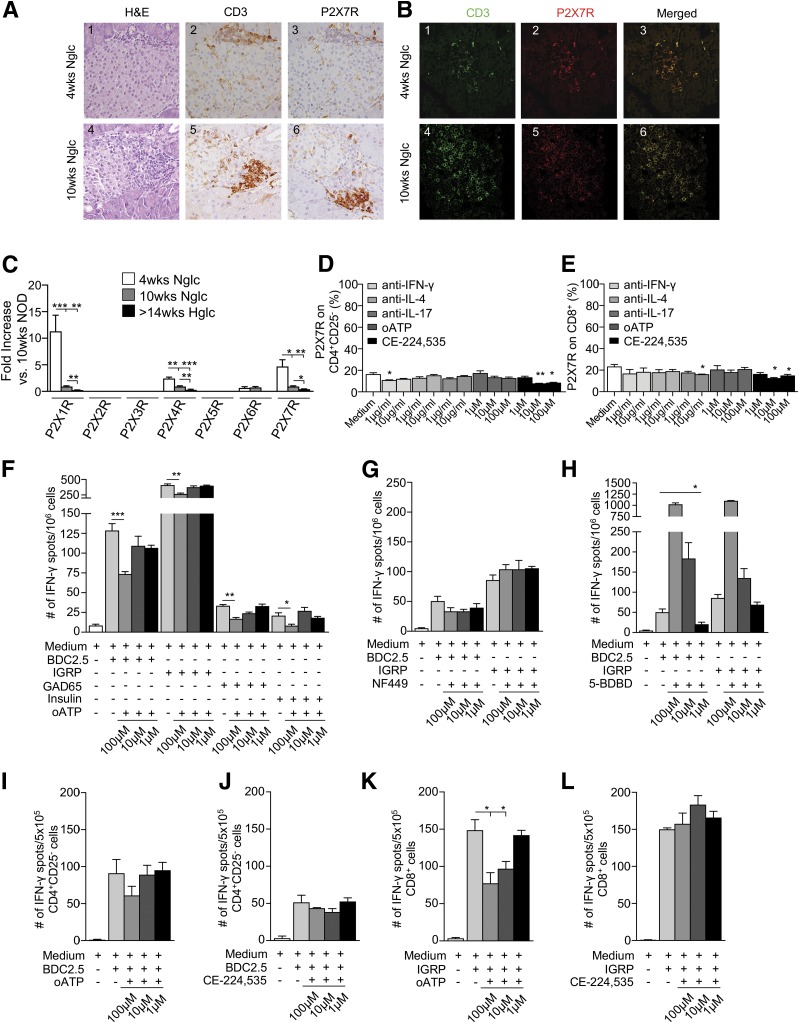
Figure 3.
P2X7R+ T cells make up the vast majority of pancreas-infiltrating cells, while P2X7R blockade reduces the autoimmune response in vitro. A and B: Islets from 10-week-old and, to a lesser extent, 4-week-old NOD mice showed CD3+ T-cell (green) infiltrate, which appeared to be predominantly P2X7R+ (red). H&E, hematoxylin and eosin; Nglc, normoglycemic. C: P2X1R, P2X4R, and P2X7R mRNA pancreatic expression was evident in 4-week-old and, to a lesser extent, 10-week-old NOD mice. Hglc, hyperglycemic. D: Significant downregulation of P2X7R expression on CD4+CD25– T cells was observed when cells were cultured with anti–IFN-γ or the P2X7R inhibitor CE-224,535 as compared with medium alone. E: P2X7R downregulation was observed on CD8+ T cells cultured with anti–IL-17 or CE-224,535 as compared with medium alone. F: Splenocytes from hyperglycemic NOD mice cultured with the CD4/CD8-restricted islet mimotope peptides BDC2.5/IGRP, respectively, or with GAD65 and insulin in the presence of oATP showed a reduction in the number of IFN-γ+ T cells as compared with controls. G and H: Splenocytes from hyperglycemic NOD mice cultured with BDC2.5 or IGRP peptides in the presence of different concentrations of the P2X1R antagonist NF449 or the P2X4R antagonist 5-BDBD did not show any effect or showed an increase in the number of IFN-γ+ T cells, respectively. I–L: oATP but not CE-224,535 reduced the number of IFN-γ+ CD8+ but not CD4+ T cells when CD4+CD25– cells isolated from BDC2.5 TCR Tg NOD mice (I and J) or CD8+ T cells isolated from 8.3 TCR Tg NOD mice (K and L) were cultured with BDC2.5 or IGRP peptides, respectively, in the presence of different concentrations of oATP or CE-224,535. Data are expressed as mean ± SEM. Data are representative of at least n = 3 mice. *P < 0.05; **P < 0.01; ***P < 0.001.

