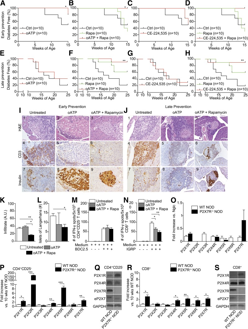
Figure 4.
P2X7R blockade delays diabetes in NOD mice and reduces islet infiltration and the autoimmune response by CD8+ T cells. A–H: The effect of P2X7R blockade was tested in early (4-week-old NOD mice) and late (10-week-old NOD mice) prevention studies in vivo using oATP, CE-224,535, and a combination of oATP or CE-224,535 with clinical-grade-dose rapamycin (Rapa). The treatment with oATP and oATP plus rapamycin (A and B), but not with CE-224,535 alone or with rapamycin (C and D), significantly delayed the onset of diabetes in the early prevention study setting as compared with untreated animals; data are representative of n = 10 mice per group. In the late prevention study, while oATP and CE-224,535 alone did not show any effect on the onset of diabetes (E and G), their combination with rapamycin successfully delayed diabetes onset (F and H); data are representative of n = 10 mice per group. I and J: Representative hematoxylin and eosin (H&E) staining and immunohistochemical analysis of pancreatic islet tissue sections from the different groups of treated and untreated NOD mice. K and L: Semiquantitative analysis of islet infiltrate in 4-week-old treated NOD mice revealed that oATP plus rapamycin is more effective in reducing islet infiltration, although it is associated with an overall reduced number of islets. A.U., arbitrary units. M and N: CD4+CD25– and CD8+ T cells isolated from the spleens of 10-week-old treated and untreated NOD mice and cultured in the presence of BDC2.5 or IGRP peptides, respectively, showed a reduced number of IFN-γ+ CD8+ but not CD4+ T cells in mice treated with oATP alone or in combination with rapamycin as compared with untreated mice. O: The pancreata of 10-week-old normoglycemic (Nglc) P2X7R−/− and WT NOD mice contain P2X7R mRNA. P: mRNA expression of P2X1R, P2X2R, P2X4R, P2X5R, and P2X7R in splenic CD4+CD25– cells was significantly increased in P2X7R−/− NOD mice as compared with WT NOD mice. Q: Western blot analysis of P2X1R, P2X4R, P2X7R, and eP2X7 (extracellular P2X7) in splenic CD4+CD25– T cells confirmed the absence of P2X7R protein in P2X7R−/− NOD mice as compared with WT NOD mice. R: mRNA expression of P2X1R, P2X4R, P2X5R, and P2X7R in splenic CD4+CD25– and in CD8+ T cells (except for P2X2R) were significantly increased in P2X7R−/− NOD mice as compared with WT NOD mice. S: Western blot analysis of P2X1R, P2X4R, and P2X7R in splenic CD8+ T cells confirming the absence of P2X7R protein in P2X7R−/− NOD mice as compared with WT NOD mice. Data are expressed as mean ± SEM. Data are representative of at least n = 3 mice. *P < 0.05; **P < 0.01; ***P < 0.001.

