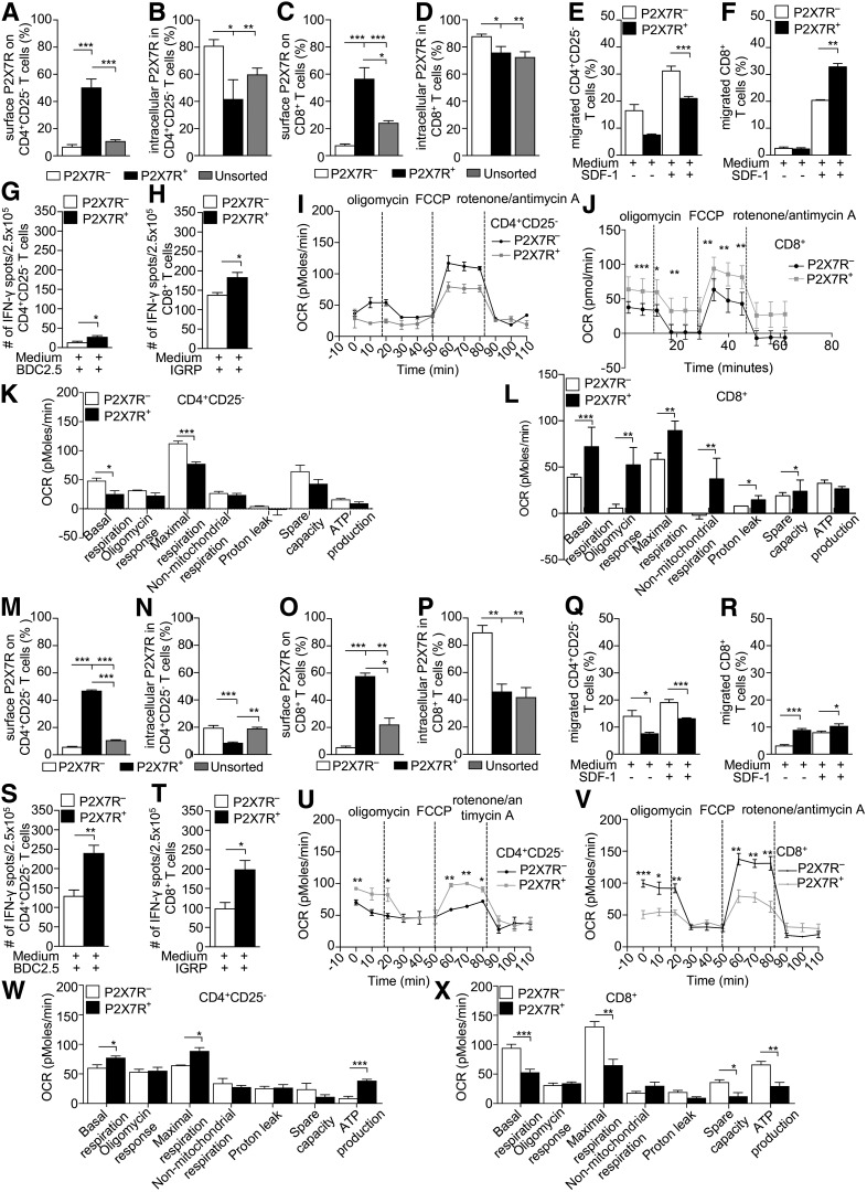
Figure 5.
Characterization of P2X7R− and P2X7R+ cells reveals dependence of CD8+ T cells on eATP/P2X7R signaling. A–D: P2X7R− CD4+CD25–/CD8+ T cells isolated from 10-week-old hyperglycemic or normoglycemic NOD or BDC2.5/8.3 TCR Tg NOD mice (n = 3 samples per group) showed no surface expression of P2X7R, while significant intracellular expression was detected. E and F: The percentage of P2X7R−CD4+CD25– T cells migrating to SDF-1 was significantly higher as compared with P2X7R+CD4+CD25– T cells, while the opposite was observed in CD8+ T cells, with a higher percentage of migrating P2X7R+CD8+ T cells as compared with P2X7R−CD8+ T cells. G and H: P2X7R+CD4+CD25–/CD8+ T cells from hyperglycemic NOD mice cultured with the CD4/CD8-restricted islet mimotope peptides BDC2.5 and IGRP, respectively, generated more IFN-γ+ T cells. I and K: Higher OCR in basal conditions and after FCCP addition was observed in P2X7R− CD4+CD25– T cells as compared with P2X7R+CD4+CD25– T cells. J and L: In P2X7R+CD8+ T cells, OCR was increased after FCCP addition and reduced in response to rotenone/antimycin A as compared with P2X7R−CD8+ T cells. While the maximal respiration capacity and SRC of P2X7R+CD8+ T cells were significantly increased, following addition of oligomycin and in response to rotenone/antimycin A, the OCR was significantly reduced as compared with P2X7R+CD8+ T cells. M–P: P2X7R−CD4+CD25–/CD8+ T cells from NOD.BDC2.5 and NOD.8.3 mice showed significant intracellular P2X7R expression as well. Q and R: Among CD4+CD25– T cells from NOD.BDC2.5 mice, the percentage of migrating P2X7R− cells was significantly higher as compared with P2X7R+ cells, while among CD8+ T cells from NOD.8.3 mice, the percentage of P2X7R+ cells migrating was higher as compared with P2X7R− cells. S and T: Among both CD4+CD25– and CD8+ T cells, the number of IFN-γ+ T cells generated during an islet peptide-based autoimmune assay was significantly higher among P2X7R+ as compared with the P2X7R− cells. U and W: Among CD4+CD25– T cells from NOD.BDC2.5 mice, an increase in OCR was observed in P2X7R+ cells in basal conditions and after FCCP injection as compared with P2X7R− cells. Among CD4+CD25– T cells from NOD.BDC2.5 mice, the basal, maximal respiration, and ATP-related OCR were significantly increased in P2X7R+ as compared with P2X7R− cells. V and X: Among CD8+ T cells from NOD.8.3 mice, OCR was increased in basal conditions and after FCCP injection in P2X7R− as compared with P2X7R+ cells. Among CD8+ T cells from NOD.8.3 mice, the basal, maximal respiration, SRC, and ATP-related OCR increased in P2X7R− as compared with P2X7R+ cells. Data are expressed as mean ± SEM. Data are representative of at least n = 3 mice. *P < 0.05; **P < 0.01; ***P < 0.001.

