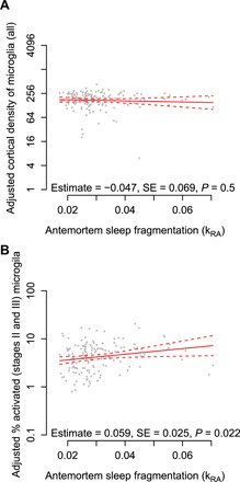
Fig. 3. Antemortem sleep fragmentation and density of cortical microglia identified by immunohistochemistry.

Average of counts in the mid-frontal and inferior temporal cortices. Partial residual plot of total microglial density (A) or proportion of stages II and II microglia (B) as a function of antemortem sleep fragmentation adjusted for age, sex, education, and postmortem interval. Each dot represents a single participant. Solid line indicates the predicted microglial count for an average participant. Dotted lines indicate 95% CIs on the prediction.
