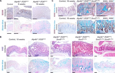
Fig. 7. Single deletion of Sox2 or Sox9 in gastric adenoma mice increases cancer severity, whereas DKO rescues tumor severity when compared to singly deleted counterparts.

(A) SOX9 immunohistochemistry (top) and Alcian blue staining (bottom) show SOX9 overexpression at sites of adenomatous epithelium in early (8 weeks) and late (18 weeks) adenoma mice (n = 3 each). Scale bars, 100 μm. (B) Corresponding H&E (top) and Alcian blue (bottom) staining shows the aberrant changes in gastric epithelial structure and cell types in the various adenoma models (n ≥ 3 for Atp4bCre;R26NICD, Atp4bCre;R26NICD;Sox2flox/flox, Atp4bCre;R26NICD;Sox9flox/flox, and Atp4bCre;R26NICD;Sox2flox/flox;Sox9flox/flox mice). Scale bars, 100 μm. (C) SOX2 immunohistochemistry in Atp4bCre;R26NICD;Sox9flox/flox mice (top) and SOX9 immunohistochemistry in Atp4bCre;R26NICD;Sox2flox/flox mice (bottom) demonstrate reciprocal activation in the absence of either SOX TF (n = 3 each). Scale bars, 100 μm.
