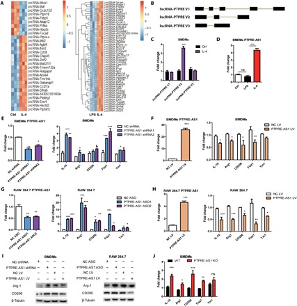
Fig. 1. PTPRE-AS1 is highly expressed and acts as a repressor in IL-4–induced M2 macrophage activation.

(A) Heatmap of antisense lncRNAs with significantly altered expression upon stimulation of BMDMs with IL-4 and LPS, respectively. (B) lncRNA-PTPRE encodes three splice variants. (C) Evaluation of the expression of three lncRNA-PTPRE splice variants in IL-4–stimulated BMDMs. (D) Expression of PTPRE-AS1 in BMDMs stimulated with IL-4 or LPS. (E) Knockdown of PTPRE-AS1 in BMDMs using two distinct shRNAs (left). (F) Overexpression of PTPRE-AS1 in BMDMs with PTPRE-AS1 LV or NC LV (left); after transfection, M2-associated gene expression in IL-4–stimulated BMDMs was quantified by RT-qPCR analysis (right). NC, negative control. (G) Knockdown of PTPRE-AS1 in RAW 264.7 cells transfected with two distinct ASOs (200 nM) (left). (H) Overexpression of PTPRE-AS1 in RAW 264.7 cells with PTPRE-AS1 LV or NC LV (left), followed by IL-4 stimulation; M2-associated gene expression was quantified by RT-qPCR (right). (I) Western blots of protein levels of Arg-1 and CD206 in BMDMs (left) and RAW 264.7 cells (right) with PTPRE-AS1 knockdown or overexpression, followed by IL-4 stimulation for 24 hours. (J) M2-associated gene expression levels in WT and PTPRE-AS1–deficient mouse BMDMs were detected by RT-qPCR after IL-4 treatment for 24 hours. Data are presented as mean ± SEM from three independent experiments. *P < 0.05; **P < 0.01; ***P < 0.001; ns, no significance.
