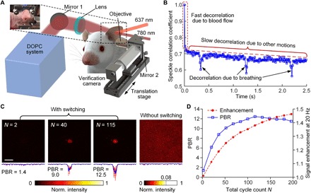
Fig. 4. In vivo demonstration of focusing light inside tumors.

(A) Schematic of the setup for focusing light inside tumors on the mouse ear in vivo. A microscope is placed on a translation stage and can be moved horizontally into the light path to image the time-reversed focus. (B) Speckle correlation coefficient as a function of time for a living mouse ear. Three speckle decorrelation characteristics were identified. (C) Normalized intensity distributions of the optical foci inside the tumor on the mouse ear. Left, with 637-nm light switching for N = 2, 40, and 115 cycles; right, without 637-nm light switching. Scale bar, 100 μm. (D) Signal enhancement of tagged photons (at 20 Hz) and the PBR of time-reversed focusing as a function of the total cycle count N.
