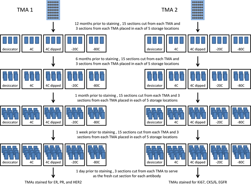
Figure 1.
Overall study design for tissue microarray (TMA) section storage. TMA 1, stained for ER, PR, and HER2, contained 60 patients that were chosen because they were positive for these markers in the patients’ medical record. TMA 2 consisted of 88 patients with triple negative breast cancer, and this TMA was stained with Cytokeratin 5/6, EGFR, and Ki67. All stored sections were compared to a fresh section that was cut one day prior to staining.

