Figure 2.
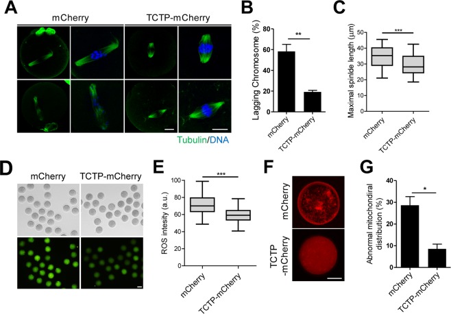
Exogenous TCTP relieves the decline of oocyte quality. Oocytes were treated with mCherry or TCTP-mCherry for 4 hours. After washing the proteins, oocytes were in vitro cultured in fresh media for 24 hours. (A–C) Spindle microtubules and chromosomes were immunostained with anti-tubulin antibody and DAPI, respectively. Representative images from three independent experiments are shown in (A). Bar, 20 μm. (B,C) Percentage of lagging chromosomes and maximal spindle length are shown. Data are expressed as means ± SEMs from three independent experiments. (D,E) Oxidative stress levels were determined by measuring fluorescence intensity following incubation with DCF-DA for 30 min. Representative images from three independent experiments are shown, along with quantification of ROS levels. Bar, 80 μm. (F,G) Mitochondrial distribution was determined by immunostaining with CytoPainter MitoRed. Representative images from three independent experiments are shown. Bar, 40 μm. The incidence of mitochondrial aggregation is shown. Data are expressed as means ± SEMs from three independent experiments. *p < 0.05, **p < 0.001, ***p < 0.0001 (Student’s t-test).

