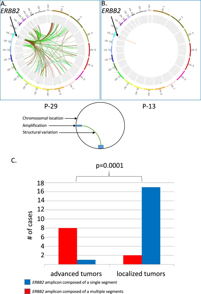
Figure 2.
ERBB2 amplicon structure is different in patient with localized and advanced disease. FAST analysis of low coverage whole genome sequencing (lcWGS) of two primary breast ER+/HER2+ tumors is visualized using Circus. The external ring represents chromosomal location; the inner ring shows areas of amplification as blue bars. Colored lines represent structural variations (SV), all the SV in an amplicon are colored with the same color. Although both (P-29 and P-13) are primary breast ER+/HER2+ tumors, P-29 was resected from a patient suffering from a metastatic disease as a palliative measure and P-13 was resected from a patient with a localized disease with curative intent. In P-29 the ERBB2 amplicon is composed of several segments (Panel A) and in P-13 of a single segment (Panel B). The ERBB2 amplicon is composed of a single segment, colored blue, in 17/19 patients with localized tumors, and is composed of multiple segments, colored red, in 8/9 patients with advanced tumors, Fisher exact test, two sided p = 0.0001 (Panel C).

