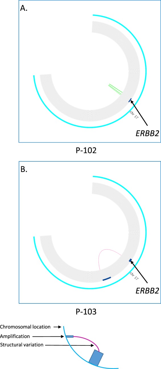
Figure 3.

Difference in ERBB2 amplicon in two breast tumors from the same patient. FAST analysis of low coverage whole genome sequencing (lcWGS) of two primary ER+/HER2+ and ER?/HER2+ breast tumors is visualized using Circus. The external ring represents chromosomal location on chromosome 17, the inner ring shows areas of amplification as blue bars. Colored lines represent structural variations (SV). Both tumors (P-102 and P-103) are primary HER2+ breast cancer. In P-102 the ERBB2 amplicon spans chr17:37,125,000-38,715,000, has eight copies and has an inverted duplication amplicon structure (panel A) while In P-103 the ERBB2 amplicon spans chr17:36,960,000-38,070,000, has 16 copies and has a double minute amplicon structure (panel B).
