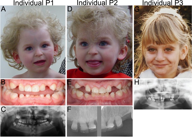
Fig. 2.
Facial appearances and dental abnormalities of the three individuals carrying biallelic POLR3GL variants. a Photograph showing the facial characteristics of individual P1 at the age of 3 years including upslanting palpebral fissures, thin lips, a flat philtrum and relatively long columella. b Intra-oral photograph of dental abnormalities of individual P1 showing an anterior open bite extending to the buccal teeth whereby only the most distal molars occlude. The central lower deciduous teeth are worn because of prolonged use. c Orthopantomographic radiograph of the full dentition of individual P1. The Orthopantomograph shows that the permanent dentition consists of the first and second molars, all other teeth are congenitally absent. In addition, it shows that the upper- and lower molar pulp chamber have a taurodontic shape. d Photograph of the face of individual P2 at the age of 3 years showing upslanting palpebral fissures, a flat philtrum and thin lips with relatively long columella. e Intraoral photograph of dental abnormalities of individual P2. Only the distal molars occlude. There is spacing in between the teeth due to the growth of the jaws and absence of permanent teeth. f Radiograph of deciduous upper frontal teeth of individual P2 showing short erratic shape of the teeth and obliterated root canals. The original shape of the crowns and the covering with composite can be seen. g Photograph showing the facial characteristics of individual P3 at the age of 8 years which include a high nasal bridge with a relatively long columella and thin lips. h Orthopantomographic radiograph of the full dentition of individual P3. Upper medial incisors and 5 molars are present in the permanent dentition with taurodontic shape of the upper- and lower molars

