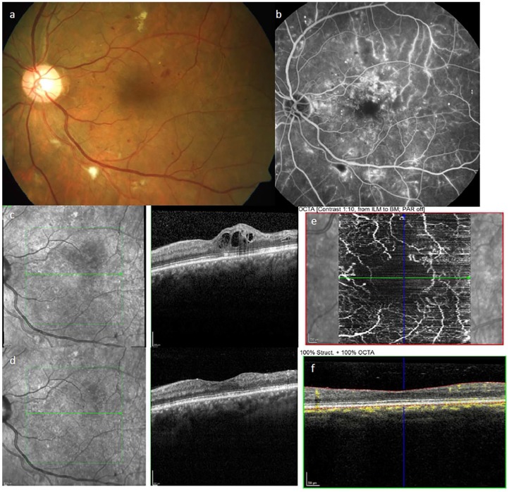
Figure 2.
Ophthalmologic findings in patients with diabetes.
Examples of pathological ophthalmologic findings in the left eye of a patient with diabetes. (a) Fundus photography showing optic nerve head and macula with signs of moderate DR: microaneurysms (red dots), small hemorrhages (red spots), hard exudates (yellow spots), and cotton-wool spots (white spots). (b) Fluorescein angiography showing the dye within the retinal vessels and capillaries, leakage of the dye outside the vessel is visible in multiple spots. (c) Spectral-domain OCT showing diabetic macula edema, the intraretinal fluid appears as black circles. (d) OCT showing near normal retinal thickness after intravitreal injection of anti-VEGF, the intraretinal fluid is almost completely resorbed. (e) OCT-angiography (without dye) showing the flow within the retinal microvessels around the central avascular arcade and (f) the corresponding OCT-angiography scan highlighting the particular segmentation of the retina (red lines) and the flow measurement (yellow) within all layers of the retina.

