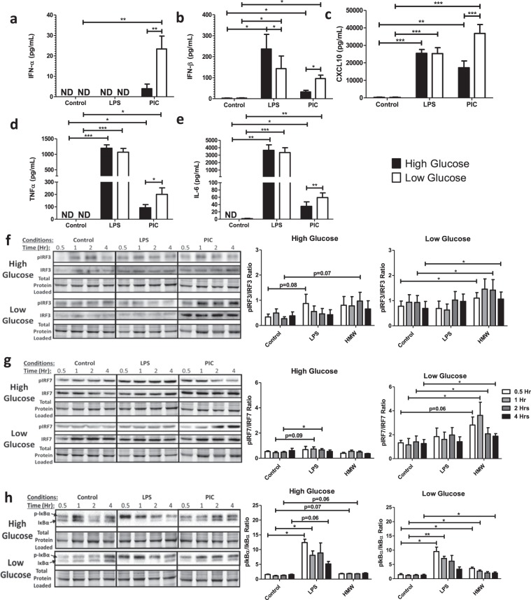
Figure 4.
Low glucose conditions are associated with increased IRF activation and increased type I IFN production. Macrophages were stimulated with either 100 ng/mL LPS or 10 ng/mL PIC for 18 hours under high glucose (25 mM) or low glucose (0.5 mM) media conditions. Supernatant was collected for assessing antiviral (IFN-α, IFN-β, CXCL10) (a–c) and pro-inflammatory (TNF-α, IL-6) cytokine (d,e) expression. Cell lysates were harvested to quantify p-IRF3 and total IRF3 (f), p-IRF7 and total IRF7 (g) and p-Iκbα and total Iκbα (h) expression via immunoblotting. Data represents mean ± SEM of four individual mice (*p < 0.05, **p < 0.01, and ***p < 0.001). For visualization purposes, the western blot images were cropped, but full-length blots and gel images can be found in Supplemental Fig. S5.

