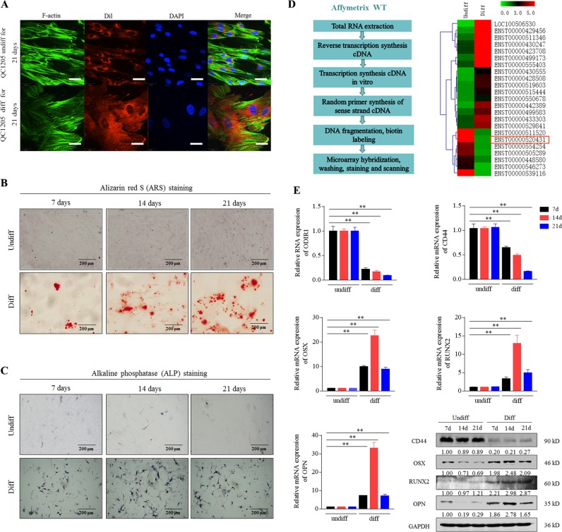
Fig. 1. Identification of regulatory lncRNAs during osteogenic differentiation of hUC-MSCs.
a The hUC-MSCs were cultured in normal growth medium (Undiff) or osteogenic differentiation medium (Diff) for 21 days, and analyzed by fluorescence laser confocal for cytoskeleton (F-actin), cell membrane (Dil) and nucleus (DAPI). Scale bar, 20 μm. b–c The diff and undiff hUC-MSCs cells were analyzed by Alizarin red S (ARS) and alkaline phosphatase (ALP) staining. Scale bar, 200 μm. d An operating flow diagram for lncRNA microarray and a heatmap for differentially expressed lncRNAs of osteogenic differentiated hUC-MSCs compared to undifferentiated hUC-MSCs. ENST00000520431 (ODIR1) was significantly decreased after osteogenic differentiation. e The hUC-MSCs were incubated for 7, 14, and 21 days, respectively, then the RNA levels of ODIR1, CD44, OSX, RUNX2, and OPN were analyzed by RT-qPCR, and the proteins levels of CD44, OSX, RUNX2, OPN were analyzed by western blots. GAPDH was used as internal controls.

