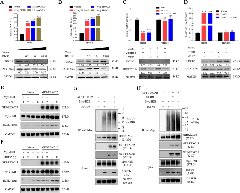
Fig. 6. ODIR1 restrains the levels of H2BK120ub protein through recruiting FBXO25.
a The hUC-MSCs were transfected with vector and different amounts (0, 0.5, 1.0, and 2.0 μg) of ODIR1 plasmid, the RNA levels of ODIR1 was measured by RT-qPCR and the proteins levels of FBXO25 and H2BK120ub were detected by western blotting. b The hUC-MSCs were transfected with vector and different amounts (0, 0.5, 1.0, and 2.0 μg) of FBXO25 plasmid, the RNA levels of FBXO25 was measured by RT-qPCR and the proteins levels of FBXO25 and H2BK120ub were detected by western blotting in cell lysates. c The hUC-MSCs were transfected with shCtrl or shODIR1 plasmid and treated with CHX, and the proteins levels of FBXO25 and H2BK120ub were analyzed by western blotting assay. d The hUC-MSCs were transfected with vector or ODIR1 plasmid and treated with MG132, and the proteins levels of FBXO25 and H2BK120ub were analyzed by western blotting assay. e The HEK293T cells were transfected with FBXO25 overexpression vector, followed by CHX treatment for indicated periods of time and then H2BK120ub were detected by western blot. f The HEK293T cells were transfected with FBXO25 overexpression vector, followed by MG132 treatment for indicated periods of time and then H2BK120ub were detected by western blot. g HEK293T cells were transfected with GFP-FBXO25, Myc-H2B and HA-Ub as indicated. After 48 h culture, the levels of ubiquitinated H2B were monitored by IP analysis, followed by western blot with indicated antibodies. h HEK293T cells were transfected with ODIR1, GFP-FBXO25, Myc-H2B, and HA-Ub as indicated. After 48 h incubation, the levels of ubiquitinated H2B were monitored by IP analysis, followed by western blot with indicated antibodies.

