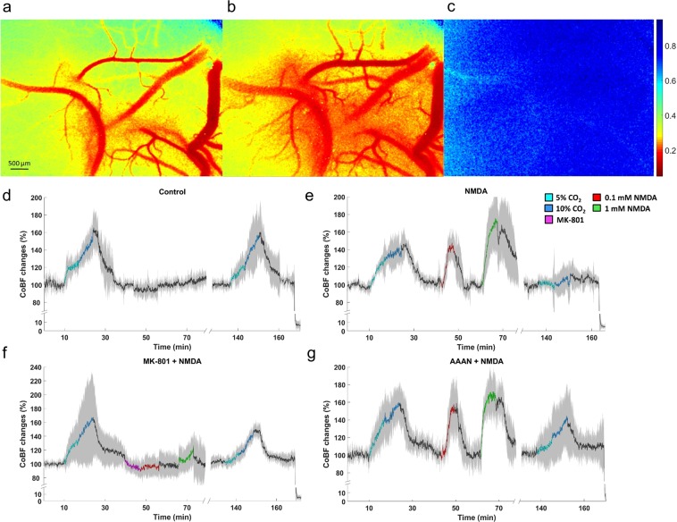
Figure 2.
Cortical blood flow (CoBF) changes to graded hypercapnia and NMDA using laser-speckle contrast imaging and analysis. (a–c) Representative LSCI contrast images obtained through the closed cranial window (a–c) with the corresponding contrast scales. Lower contrast values represent higher flow velocity in the cortical microcirculation. (a) baseline condition; (b) NMDA (1 mM) showing pial arteriolar vasodilation and increased parenchymal flow as well. (c) biological zero after euthanasia characterized by high and stable speckle contrast values marking the disappearance of the perfusion. (d–g) Summarized recordings of individual experiments, colored/black lines represent the group mean values during/between stimuli, the gray area represent the SD. (d) in the control group graded hypercapnia resulted in concentration-dependent repeatable increases in CoBF relative to the baseline. (e) Both doses of NMDA reversibly elevated CoBF, however, the CoBF response to graded hypercapnia was virtually absent after NMDA. (f) Pre- and co-treatment of MK-801 with NMDA abolished the CoBF response to NMDA and prevented the attenuation of the CoBF response to graded hypercapnia. (g) AAAN did not affect the CoBF response to hypercapnia or NMDA, however, at least partially prevented the attenuation of the microvascular response to graded hypercapnia by NMDA.

