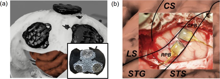
Fig. 7. The anatomical structure of the cortex guided posistions of MEA on parabelt.
a A 3D model of the skull, brain, and anchoring metal footplates (constructed by merging MRI and CT imaging). Also, a titanium mesh on a 3D-printed skull model. b Photo of the exposed area of the auditory cortex with labels added for relevant cortical structures (CS, central sulcus; LS, lateral sulcus; STG, superior temporal gyrus; STS, superior temporal sulcus; RPB, rostral parabelt; CPB, caudal parabelt).

