Figure 6.
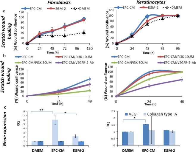
Effect of endothelial progenitor cells conditioned medium (EPC-CM) on gingival fibroblasts and keratinocytes scratch wound assay and associated genes. (a) Scratch wound assay: Cells were cultured in 10 µM zoledronic acid (ZOL) for 30 h. Immediately after scratch, the medium was replaced by fresh drug-free medium: Dulbecco’s modified Eagle medium (DMEM), EPC-CM, and endothelial growth medium without EPC secretome (EGM-2). Confluence of the wound was followed and measured automatically, P ≤ 0.038, EPC-CM vs. DMEM + drugs for gingival fibroblasts and keratinocytes. (b) Scratch wound healing assay with VEGF pathway blockage: Cells were cultured in 10 µM zoledronic acid (ZOL) for 30 h. Immediately after scratch, the medium was replaced by fresh drug-free medium: EPC-CM(+)/50 µM PI3K, EPC-CM(+)/10 µM PI3K or EPC-CM(+)/0.2 mg/ml VEGFR-2 antibody. (c) Wound healing associated genes: Cells were cultured in 10 µM zoledronic acid (ZOL) for 30 h, followed by 48 h in: DMEM, EPC-CM, or EGM-2. The mRNA of VEGFA and COL1A1 were quantified using RT-PCR. Relative quantification was calculated relative to HPRT and cell cultured in10 µM ZOL for 30 h followed by DMEM for 48 h. *P ≤ 0.05, **P ≤ 0.001. Experiments repeated tree times in triplicates.

