Figure 1.
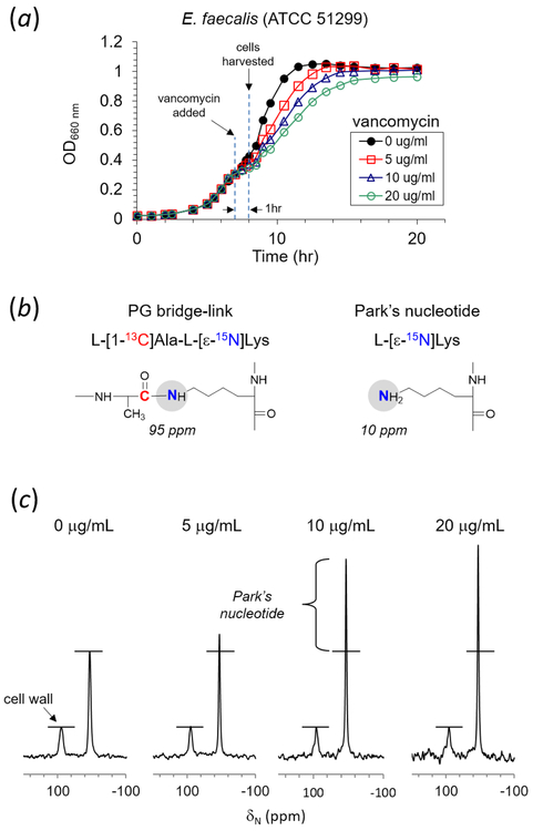
(a) Growth curves of vancomycin resistant E. faecalis (ATCC 51299) in ESM containing l-[1-13C]Ala and l-[ε-15N]Lys. Growth was monitored by measuring optical density at 660 nm (OD660nm). Vancomycin was added to final concentrations of 0, 5, 10, and 20 μg/mL during growth at OD660nm 0.3. Cells were harvested at OD660nm 0.4 for 15N-CPMAS NMR analysis. (b) Incorporation of l-[ε-15N]Lys into the PG bridge-link is visible as a lysyl-amide at 95 ppm, and into Park’s nucleotide as a lysyl-amine at 10 ppm in 15N-CPMAS spectra. (c) 15N-CPMAS spectra of E. faecalis labeled with l-[1-13C]Ala and l-[ε-15N]Lys. Spectra are normalized to 95-ppm intensity. l-[ε-15N]Lys incorporated into PG bridge-linked resonates at 95 ppm, and l-[ε-15N]Lys into proteins and Park’s nucleotide at 10 ppm. VRE grown in presence of vancomycin show Park’s nucleotide accumulation, indicating that vancomycin inhibited the transglycosylation step of PG biosynthesis. 15N-CPMAS spectra of VRE grown in vancomycin concentrations of 0, 5, 10, and 20 μg/mL are results of 74092, 80000, 70512, and 80000 accumulated scans, respectively. The magic angle spinning was at 5000 Hz. All measurements were carried out at ambient room temperature.

