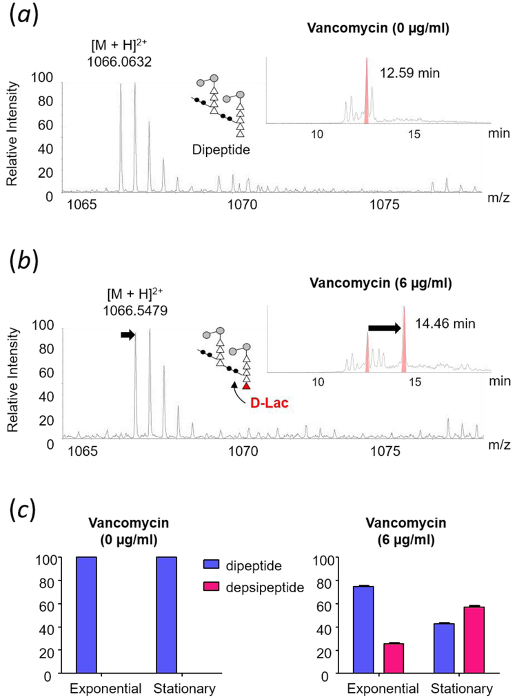
Figure 4.
Mass spectra of doubly-charged PG dimers identified from mutanolysin-digested isolated cell wall of vancomycin-resistant E. faecalis grown in absence (a) and presence of vancomycin (b) at stationary growth phase. The chemical structure, formula, and exact mass for PG dimers are provided in Supplementary Fig. S1. Select ion chromatograms (inset) show that D-Ala-D-Lac substituted PG dimer has a longer retention time that resolves it from the unmodified dimer. (c) Changes in the PG composition of dipeptide and depsipeptide terminated PG stems in the cell wall of E. faecalis grown in absence (left) and presence of vancomycin (right) during exponential and stationary growth phases. Muropeptides with PG stems terminating in d-Ala-d-Lac are only found in the cell wall of VRE grown in presence of vancomycin. Error bars represent 95% confidence interval.

