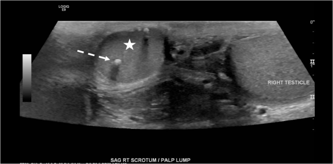
Fig. 1.
Sagittal greyscale ultrasound image shows a well-circumscribed, oval-shaped mass (white star) superior to the right testicle (labeled). The mass is isoechoic to the right testicle with similar echotexture. An echogenic, shadowing calcification (white dashed arrow) is noted within the supratesticular mass.

