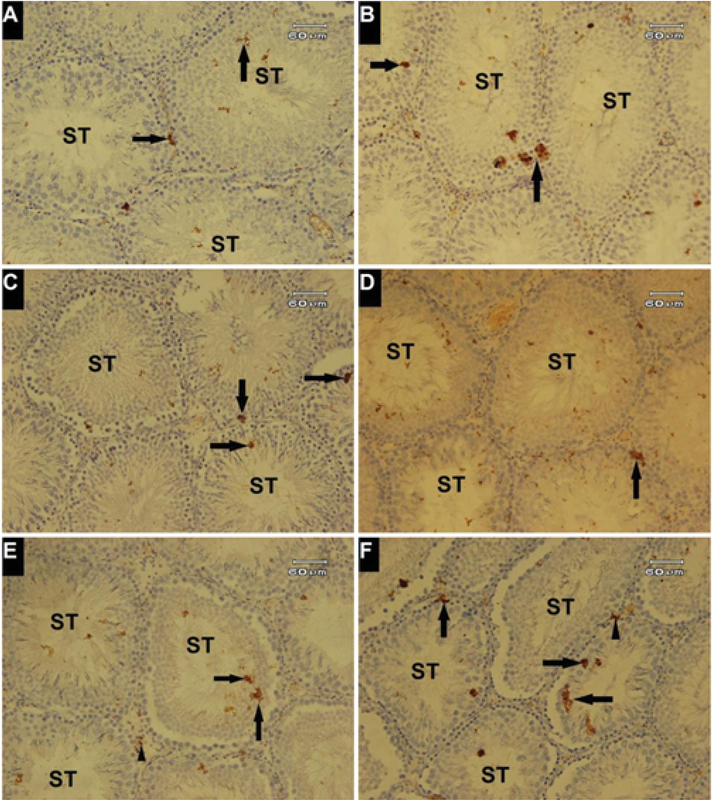
Figure 4.

Expression of p53 protein in testicular tissue. Immunohistochemical study. (A) Faint positive reaction observed (Arrows) in seminiferous tubules (ST); (B) positive reaction to p53 expression (Arrows) in PTX-treated group; (C&D) p53 positive reaction in germinal epithelium (Arrows) in the PTX + MSG30 and PTX + MSG60 groups; (E&F) Increment of positive reaction was observed in germinal epithelium (Arrows) with positive reactivity of interstitial connective tissue (Arrowheads) in the MSG30 + PTX and MSG60 + PTX groups. Magnification 200.
