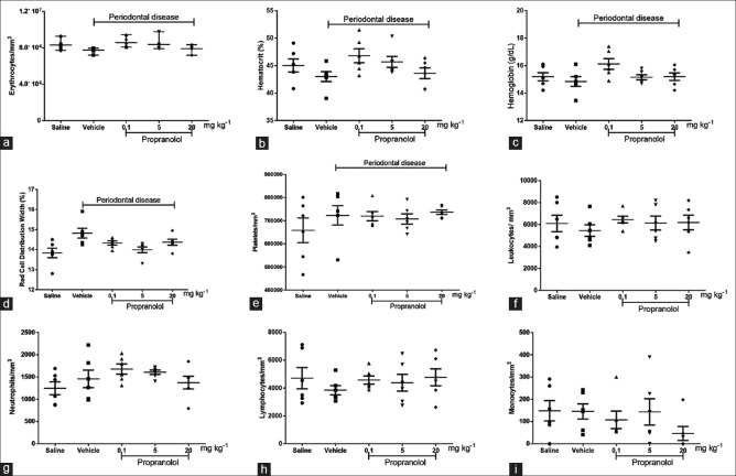
Figure 3.
Evaluation of hematological parameters. As depicted above, erythrocytes (a), hematocrit (b), hemoglobin (c), red cell distribution width (d), platelets (e), total leukocytes (f), neutrophils (g), lymphocytes (h), and monocytes (i) were quantified. The erythrocytes, hematocrit, hemoglobin, and red cell distribution width were evaluated automatically (ABX MICROS 60, Horiba ABX Diagnostics; France). The total number of leukocytes was counted with a Neubauer chamber, and the differential cell count (100 cells in total) was obtained from the stained blood smears. ANOVA and Bonferroni's test were used for group comparison

