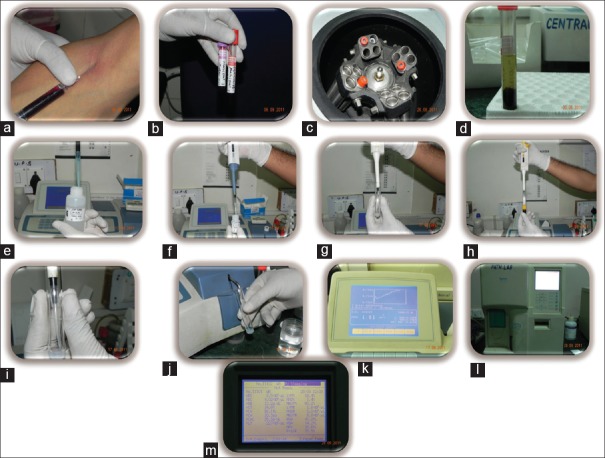
Figure 3.
(a) Blood draw, (b) Blood separated for haemetological and serological analysis, (c) Cetrifiguation for serum separation, (d) Serum separated, (e, f, g) Reagents 1&2 for CRP analysis mixed in tube, (h, i) Serum aspirated and added to reagent mix, (j) Sample loaded for CRP analysis, (k) CRP levels displayed as graph and values on machine display, (l) Automated Haematology analyzer. (m) Haemetological counts displayed on analyzer

