Figure 17.

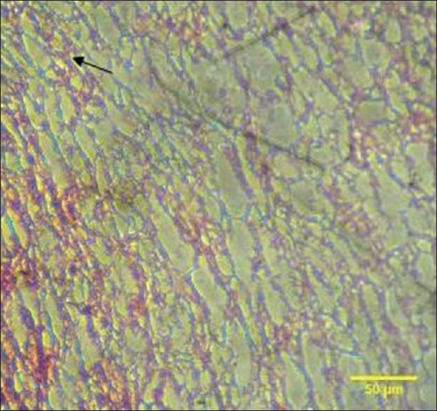
The arrow shows light microscopic analysis of platelet-rich fibrin clot showing a highly organized network of fibers. The fibrin is prominent and highly distinguishable. Dense cellular components are also observed

The arrow shows light microscopic analysis of platelet-rich fibrin clot showing a highly organized network of fibers. The fibrin is prominent and highly distinguishable. Dense cellular components are also observed