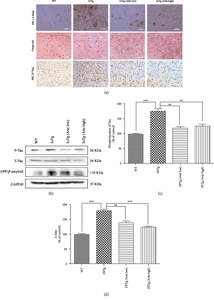
Figure 9.
Artemether treatment reduced Aβ deposition and phosphorylation of Tau in the brain cortex of 3xTg-AD mice. Artemether was administered to mice by intraperitoneal injection, once a day, at low doses of 5 mg/kg and high doses of 20 mg/kg, for 4 weeks. Thereafter, the brain cortex of wild-type (WT) compared to that of 3xTg-AD mice treated with either a low (Arte low) or high dose (Arte high) of Artemether or untreated (3xTg). (a) Immunohistochemistry of amyloid-β, Congo red staining (label amyloidosis), and phosphorylated tau (scale bar = 50 μm). Brain cortex area analyzed for amyloid-β, Congo red staining, and phosphorylated tau was the same as before (Figure 7); five slides per mouse and five mice per treatment group were used for analysis. (b) Western blot of β-amyloid and phosphorylation of Tau in each animal group; three mice per treatment group were used for western blot. (c, d) Quantitation of western blots. ∗∗p < 0.01 and ∗∗∗p < 0.001 were considered significantly different.

