Figure 3.
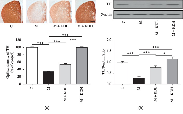
Tyrosine hydroxylase (TH) immunoreactivity and TH expression in the striatum. (a) TH-positive dopaminergic nerve fibers in the striatum (scale bar; 200 μm) and optical density of TH-positive dopaminergic nerve fibers in the striatum. (b) Expression of TH in the striatum by western blot and quantification of TH expression relative to β-actin for each group. ∗p < 0.05 and ∗∗∗p < 0.001 by one-way ANOVA followed by the Newman–Keuls post hoc test (n = 4 for each group).

