
Fig. 5.
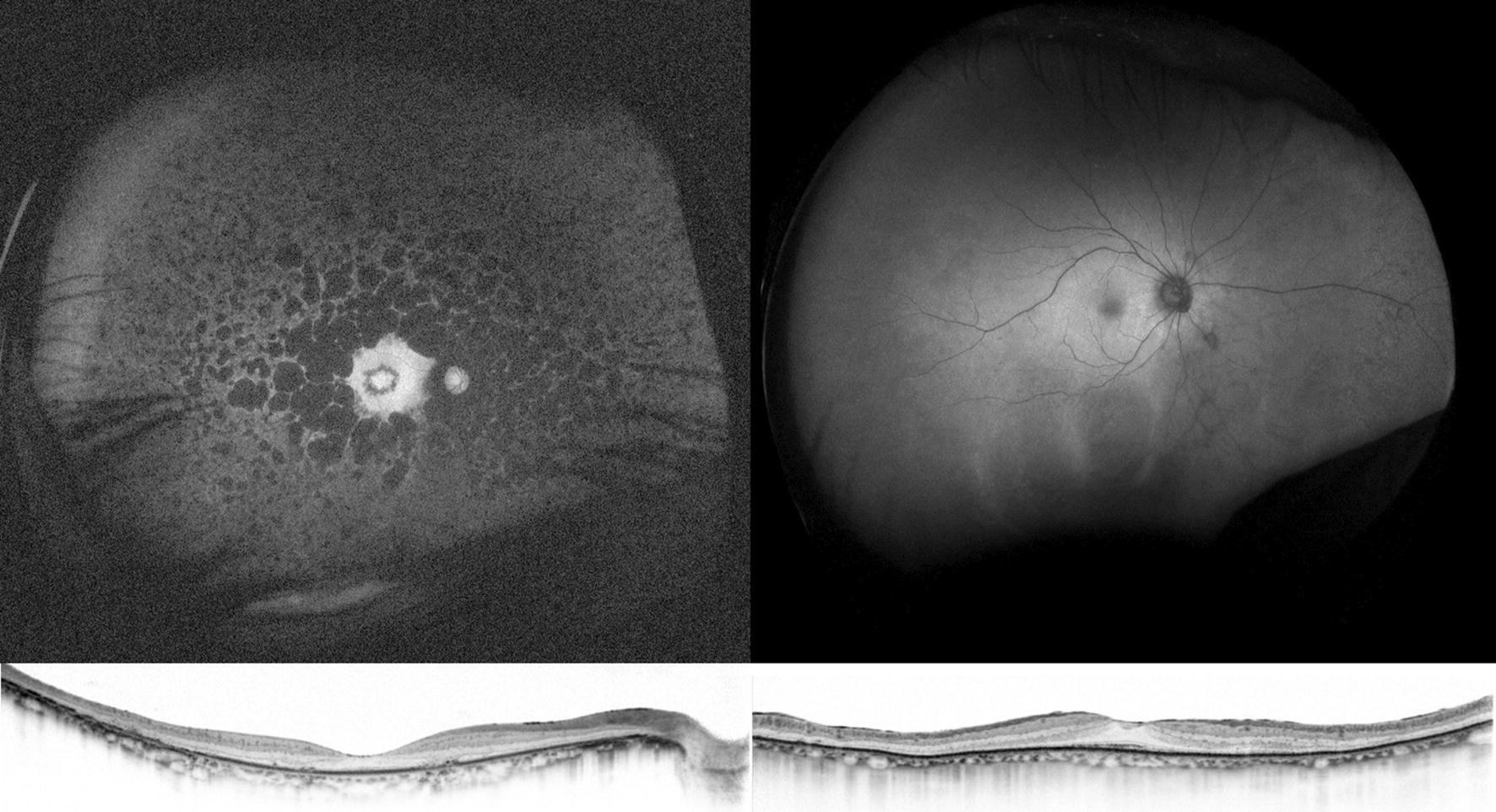
Optical coherence tomography and ultra-widefield fundus autofluorescence (FAF) in a 59-year-old patient with retinitis pigmentosa (RP; left) and an 82-year-old patient with subacute vision loss and night blindness (right). Optical coherence tomography revealed thinning of the paracentral outer nuclear layer and loss of the ellipsoid zone in these two patients. However, ultra-widefield FAF shows minimal changes in the patient on the right compared to the patient on the left. In RP, development of a granular and patchy area of decreased FAF depends on patient age and duration of the disease [43]. Thus, it is very unlikely that a patient with RP in the ninth decade of life would show almost normal FAF. The patient was screened for a systemic tumor, and lung cancer was detected. The patient was finally diagnosed with cancer-associated retinopathy