
Fig. 6.
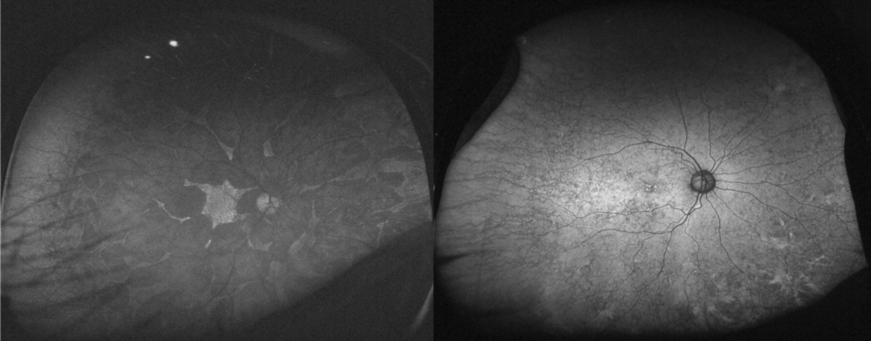
Ultra-widefield fundus autofluorescence (FAF) images of a patient with choroideremia (left) and his mother (right). Patients with choroideremia show patchy or granular areas of decreased FAF throughout the retina. Large choroidal vessels are sometimes visible in the decreased FAF area. Interestingly, an asymptomatic female carrier may show an abnormal reticular or mottled pattern of FAF. This finding may help in the diagnosis