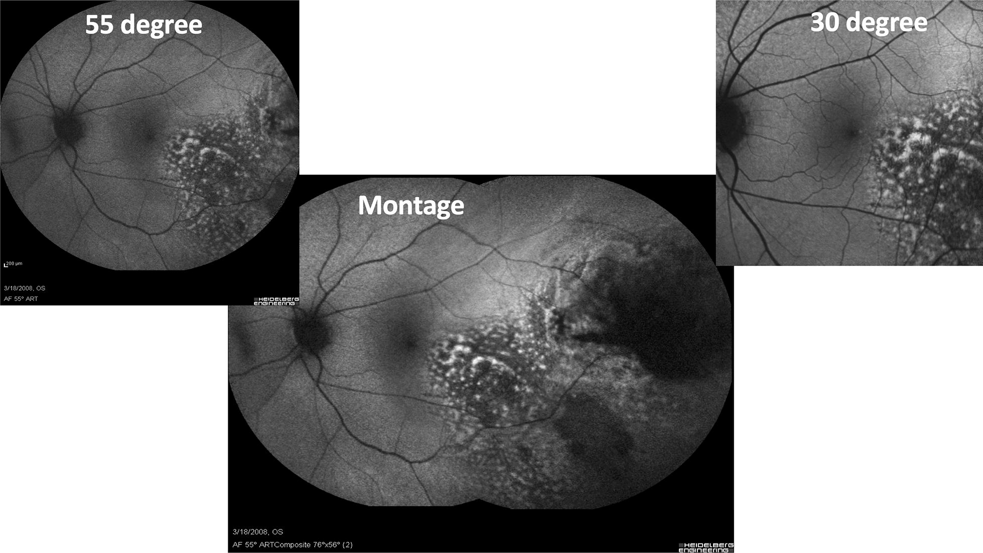
Fig. 2.

Fundus autofluorescence of a choroidal melanoma with the Heidelberg Spectralis (Heidelberg, Engineering, Inc., Heidlberg, Germany) using a standard 30° image (upper right), an extended 55° image (upper left), and a montage image (center). The choroidal melanoma demonstrates increase hyperautofluorescense corresponding to clinical areas of orange pigment as well as RPE changes
