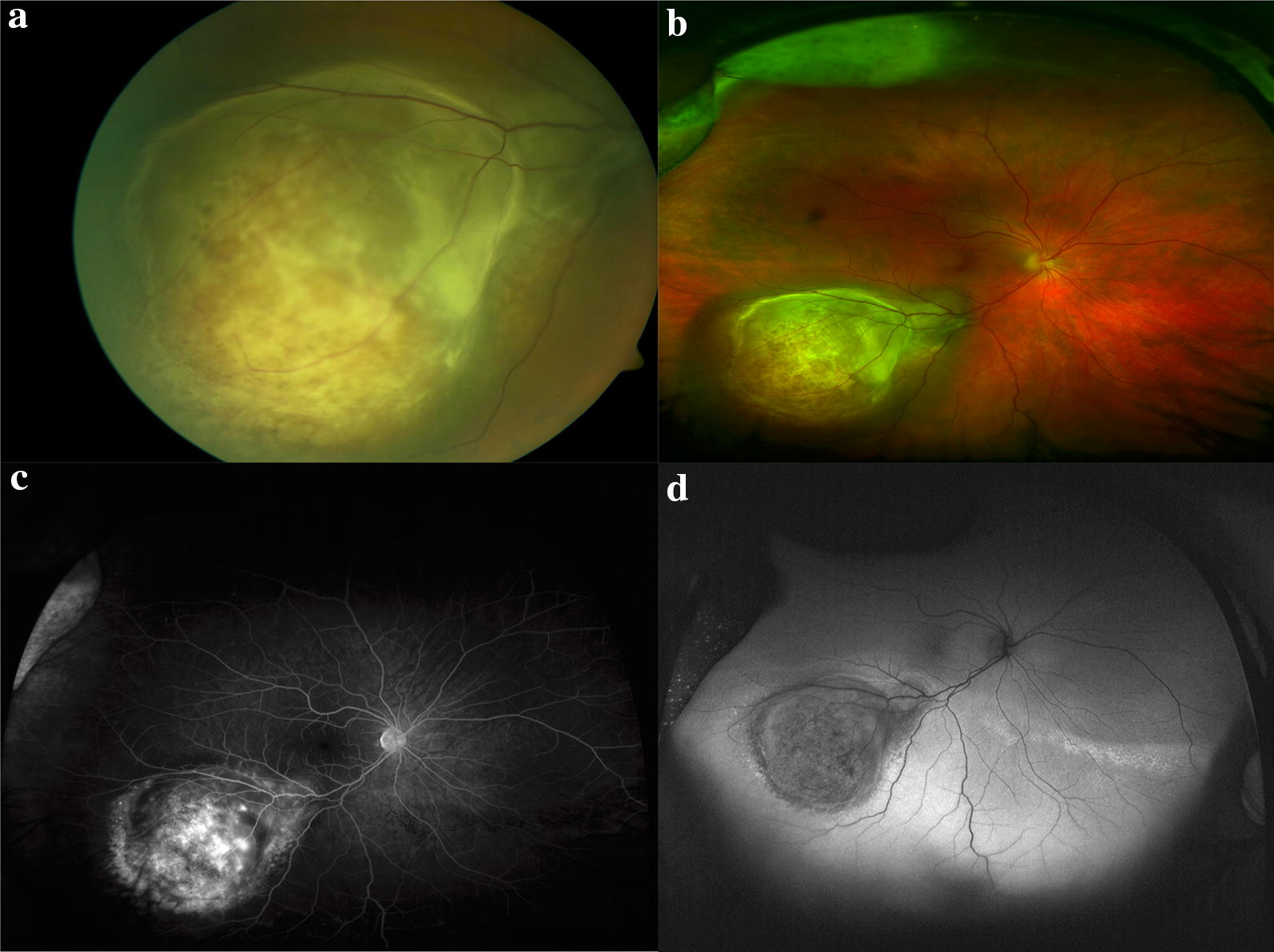
Fig. 7.

multimodal imaging of uveal melanoma. a Standard fundus photography demonstrating a choroidal mass with overlying retinal pigmentary changes and small area of detachment and subretinal fluid. b Optos wide-field imaging fo the same mass capturing the peripheral margins of the tumor. c Optos fluorescein angiography demonstrates irregular filling of the mass without other lesions visible in the retina. d Wide-field fundus autofluorescence of the tumor reveals hypo autofluorescence of the mass with inferior hyperautofluorescence consistent with subretinal fluid on examination
