Fig. 8.

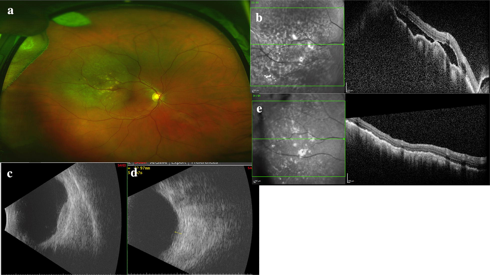
Multimodal imaging of a uveal metastasis from lung adenocarcinoma. The patient presented with reduced vision in his right eye and clinical examination revealed a large, placoid-like mass at the superotemporal arcade with overlying pigmentary changes and associated fluid. a Optos wide-field imaging of the placoid lesion with overlying pigmentary changes. b OCT with en face image with pigmentary changes, irregular, low-lying choroidal mass and resulting undulations of the choroid and associated subretinal fluid. c Ultrasonography reveals the lesion is irregular. d After target treatment the lesion thickness decreases on ultrasonography and e the choroidal mass has resolved and the subretinal fluid absorbed
