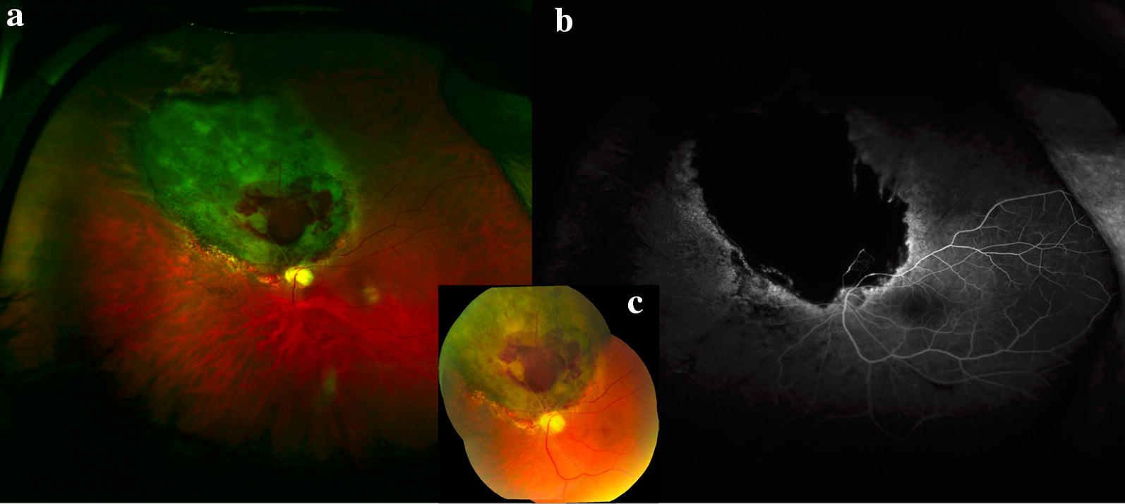
Fig. 9.

Radiation retinopathy following melanoma treatment. a Optos wide-field image of radiation retinopathy following melanoma treatment with brachytherapy. Superonasal to the disc is a flat chorioretinal scar with retinal hemorrhage and adjacent pigmentary changes. b Wide-field Optos fluorescein angiography reveals nonperfusion in the area of the scar with late leakage at the border with early development of radiation retinopathy. c Montage fundus photo demonstrating superonasal hemorrhage and radiation retinopathy
