Fig. 6.
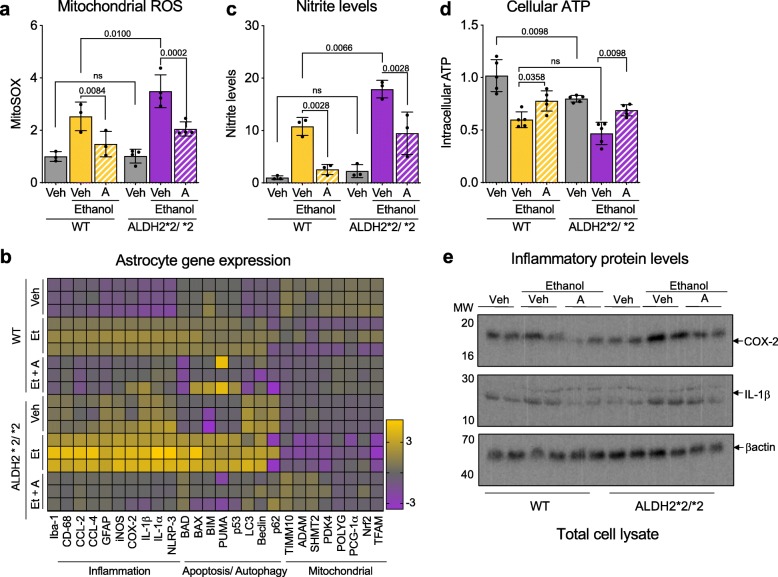
Cultured primary astrocyte derived from ALDH2*2/*2 mice are more activated in response to ethanol relative to primary astrocytes of WT mice. a) Measurement of mitochondrial ROS using MitoSOX™ in primary astrocytes in the presence or absence of Alda-1 (20 μM/24 h; 50 mM Ethanol); Veh – Vehicle; A – Alda-1. b) Heat map representation of qPCR analysis of genes associated with inflammation, apoptosis, autophagy and mitochondrial health; Veh – Vehicle; A – Alda-1. c) Nitrite levels were determined in primary astrocytes using Griess reagent kit in the presence or absence of Alda-1 (20 μM/24 h; 50 mM Ethanol); Veh – Vehicle; A – Alda-1. d) Measurement of cellular ATP levels using CellTiter-Glo Luminescent Cell Viability kit in primary astrocytes in the presence or absence of Alda-1 (20 μM/24 h; 50 mM Ethanol); Veh – Vehicle; A – Alda-1. e) Levels of cellular COX-2 and interleukin-1β release at 6 h were determined by immunoblotting in primary astrocytes in the presence or absence of Alda-1 (20 μM; 50 mM Ethanol). β-actin was used as loading control; Veh – Vehicle; A – Alda-1. Data information: Mean, standard deviation, and p-values are shown. Results are presented as fold of control. n = 3–4 independent biological replicates; probability by one-way ANOVA (with Holm-Sidak post hoc test)

