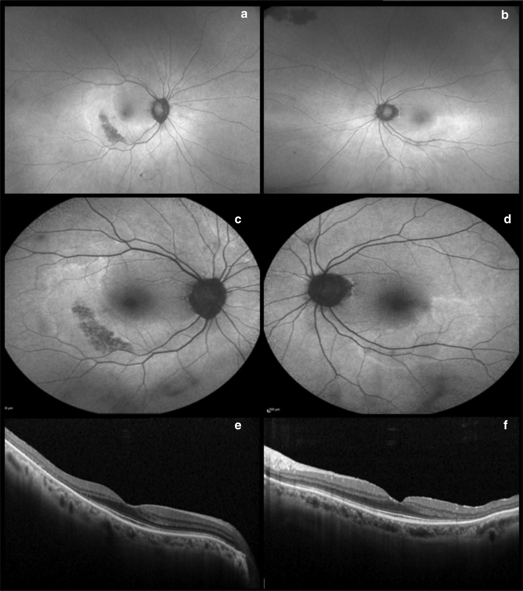
Fig. 2.

Hydroxychloroquine (Plaquenil). Optos ultra-widefield (a and b) and Heidelberg fundus autofluorescence (c and d) illustrate a more eccentric pericentral hyperautofluorescent ring corresponding to photoreceptor atrophy, sparing the fovea. Spectral domain-OCT displays bilateral and severe inner segment ellipsoid zone loss in the temporal perifovea (e and f)
