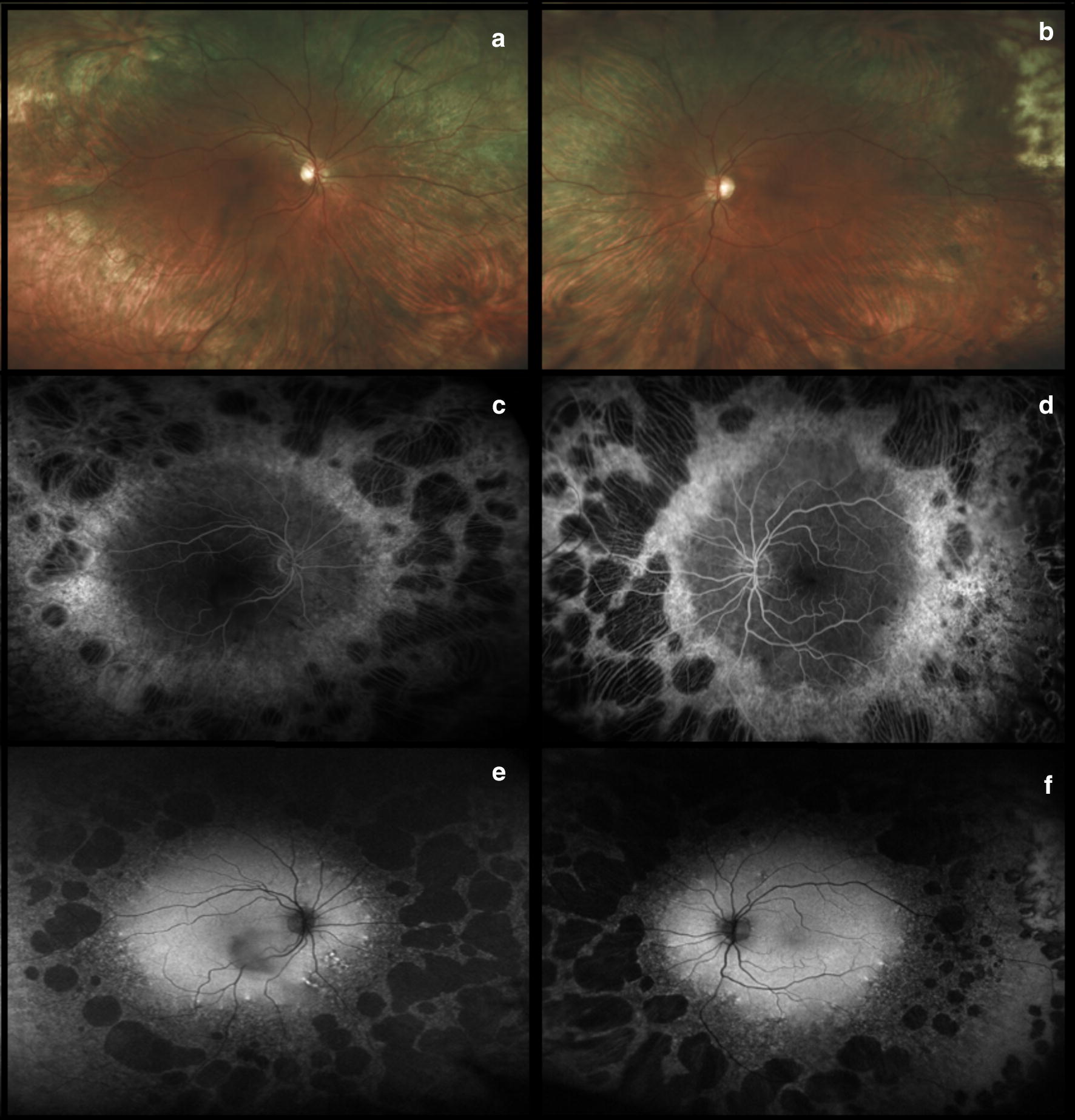
Fig. 5.

Didanosine. Ultra-widefield fundus photographs illustrate diffuse peripheral chorioretinal atrophy and sparing of the posterior pole (a and b). This atrophy is confirmed by the presence of large nummular bilateral areas of geographic atrophy on fluorescein angiography (c and d) and fundus autofluorescence (e and f). Images provided courtesy of Scott R Sneed M.D. and through permission from Haug SJ, Wong RW, Day S, Choudhry N, Sneed S, Prasad P, Read S, McDonald RH, Agarwal A, Davis J, Sarraf D. Didanosine retinal toxicity. Retina 2016;36 Suppl 1:S159-S167
