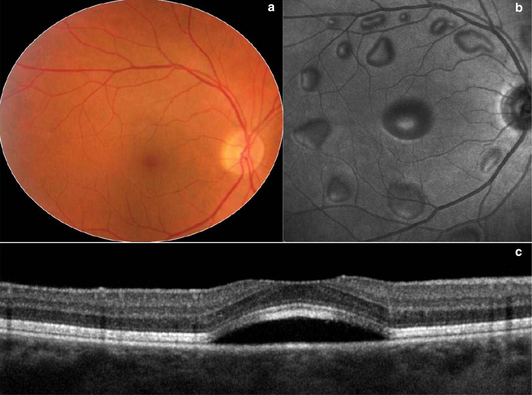
Fig. 7.

MEK inhibitor. Wide angle color fundus photograph (a) and en-face near infra-red image (b) of the right eye from a patient treated with MEK inhibitor illustrates multiple serous retinal detachments. Spectral domain-OCT (c) through the fovea in an unrelated patient displays the characteristic sub-foveal serous retinal detachment due to MEK inhibitor toxicity. Images provided courtesy of Giuseppe Querques M.D. and Enrico Borrelli M.D.
