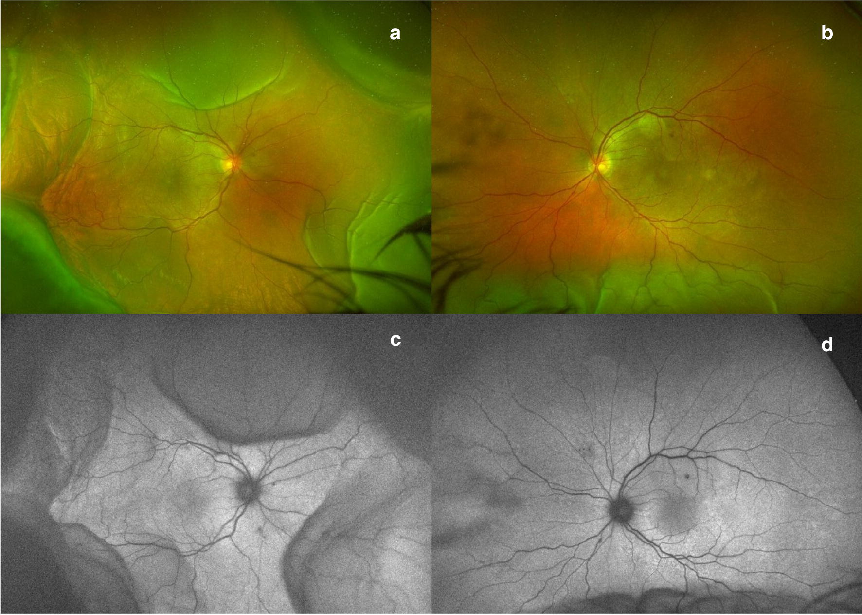
Fig. 8.

Checkpoint inhibitors. Ultra-widefield color fundus photograph (a and b) and fundus autofluorescence (c and d) from a patient with combination checkpoint inhibitor therapy (ipilimumab and nivolumab) illustrates ciliochoroidal effusions greater in the right than left eye. Images provided courtesy of Edmund Tsui M.D. and Yasha Modi M.D.
