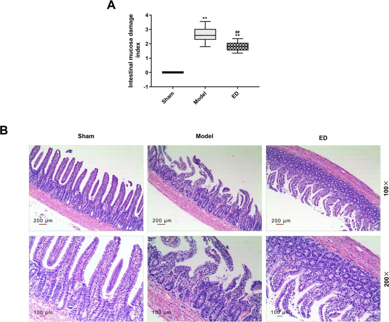
Fig. 1.
The change of intestinal mucosal tissue of rats after burn injury was observed. a The intestinal mucosa damage index was measured by colon mucosal damage index. b The pathological change of intestinal mucosal tissue was observed by using hematoxylin-eosin staining. A total of 60 Wistar rats (half male and half female) were divided into 5 groups, namely, sham group (25 °C water, n = 10), model group (100 °C water, n = 10), ED group (edaravone, 100 °C water, n = 20), scrambled group (100 °C water, n = 10) and antago group (100 °C water, n = 10). The rats were exposed to 25 °C or 100 °C water for 15 s after anesthesia (intraperitoneal injection of 50 mg/kg sodium pentobarbital). The ED group, scrambled group and antago group were given intraperitoneal injection of 9 mg/kg ED. Then the scrambled group was given intraperitoneal injection of 2 μg scrambled antagomir, and antago group was given intraperitoneal injection of 2 μg antagomiR-320. All rats were anesthetized and euthanized. **P < 0.01 vs. Sham, ##P < 0.01 vs. Model

